
Pregnancy is typically broken into three periods, or trimesters, each of about three months.[29] While there are no hard and fast rules, these distinctions are useful in describing the changes that take place over time.
First trimester
Traditionally, doctors have measured pregnancy from a number of convenient points, including the day of last menstruation, ovulation, fertilization, implantation and chemical detection. In medicine, pregnancy is often defined as beginning when the developing embryo becomes implanted into the endometrial lining of a woman's uterus. In some cases where complications may have arisen, the fertilized egg might implant itself in the fallopian tubes, the cervix, the ovary or in the abdomen, causing an ectopic pregnancy. In the case of an ectopic pregnancy, there is no way for the pregnancy to progress normally. If left untreated, it can cause harm and possibly death for the mother when a rupture occurs. Sometimes it will go away on its own, but otherwise a surgical procedure or medicine is given to remove the tubal pregnancy, since there is no way of the pregnancy being able to continue safely. Most pregnant women do not have any specific signs or symptoms of implantation, although it is not uncommon to experience minimal bleeding at implantation. Some women will also experience cramping during their first trimester. This is usually of no concern, unless there is spotting or bleeding as well. After implantation, the uterine endometrium is called the decidua. The placenta, which is formed partly from the decidua and partly from outer layers of the embryo, connects the developing fetus to the uterine wall to allow nutrient uptake, waste elimination, and gas exchange via the mother's blood supply. The umbilical cord is the connecting cord from the embryo or fetus to the placenta. The developing embryo undergoes tremendous growth and changes during the process of fetal development.
Morning sickness occurs in about seventy percent of all pregnant women, and typically improves after the first trimester. Although described as "morning sickness", women can experience this nausea during afternoon, evening, and throughout the entire day.
In the first 12 weeks of pregnancy, the nipples and areolas darken due to a temporary increase in hormones.
The first 12 weeks of pregnancy are considered to make up the first trimester. The first two weeks from the first trimester are calculated as the first two weeks of pregnancy even though the pregnancy does not actually exist. These two weeks are the two weeks before conception and include the woman's last period.
The third week is the week in which fertilization occurs and the 4th week is the period when implantation takes place. In the 4th week, the fecundated egg reaches the uterus and burrows into its wall which provides it with the nutrients it needs. At this point, the zygote becomes a blastocyst and the placenta starts to form. Moreover, most of the pregnancy tests may detect a pregnancy beginning with this week.
The 5th week marks the start of the embryonic period. This is when the embryo's brain, spinal cord, heart and other organs begin to form. At this point the embryo is made up of three layers, of which the top one (called the ectoderm) will give rise to the embryo's outermost layer of skin, central and peripheral nervous systems, eyes, inner ear, and many connective tissues. The heart and the beginning of the circulatory system as well as the bones, muscles and kidneys are made up from the mesoderm (the middle layer). The inner layer of the embryo will serve as the starting point for the development of the lungs, intestine and bladder. This layer is referred to as the endoderm. An embryo at 5 weeks is normally between 1⁄16 and 1⁄8 inch (1.6 and 3.2 mm) in length.
In the 6th week, the embryo will be developing basic facial features and its arms and legs start to grow. At this point, the embryo is usually no longer than 1⁄6 to 1⁄4 inch (4.2 to 6.3 mm). In the following week, the brain, face and arms and legs quickly develop. In the 8th week, the embryo starts moving and in the next 3 weeks, the embryo's toes, neck and genitals develop as well. According to the American Pregnancy Association, by the end of the first trimester, the fetus will be about 3 inches (76 mm) long and will weigh approximately 1 ounce (28 g).
Second trimester
Weeks 13 to 28 of the pregnancy are called the second trimester. Most women feel more energized in this period, and begin to put on weight as the symptoms of morning sickness subside and eventually fade away.
In the 20th week, the uterus, the muscular organ that holds the developing fetus, can expand up to 20 times its normal size during pregnancy. Although the fetus begins to move and takes a recognizable human shape during the first trimester, it is not until the second trimester that movement of the fetus, often referred to as "quickening", can be felt. This typically happens in the fourth month, more specifically in the 20th to 21st week, or by the 19th week if the woman has been pregnant before. However, it is not uncommon for some women not to feel the fetus move until much later. The placenta fully functions at this time and the fetus makes insulin and urinates. The reproductive organs distinguish the fetus as male or female.
Third trimester

Final weight gain takes place, which is the most weight gain throughout the pregnancy. The fetus will be growing the most rapidly during this stage, gaining up to 28 g per day. The woman's belly will transform in shape as the belly drops due to the fetus turning in a downward position ready for birth. During the second trimester, the woman's belly would have been very upright, whereas in the third trimester it will drop down quite low, and the woman will be able to lift her belly up and down. The fetus begins to move regularly, and is felt by the woman. Fetal movement can become quite strong and be disruptive to the woman. The woman's navel will sometimes become convex, "popping" out, due to her expanding abdomen. This period of her pregnancy can be uncomfortable, causing symptoms like weak bladder control and backache. Movement of the fetus becomes stronger and more frequent and via improved brain, eye, and muscle function the fetus is prepared for ex utero viability. The woman can feel the fetus "rolling" and it may cause pain or discomfort when it is near the woman's ribs and spine.
There is head engagement in the third trimester, that is, the fetal head descends into the pelvic cavity so that only a small part (or none) of it can be felt abdominally. The perenium and cervix are further flattened and the head may be felt vaginally. Head engagement is known colloquially as the baby drop, and in natural medicine as the lightening because of the release of pressure on the upper abdomen and renewed ease in breathing. However, it severely reduces bladder capacity, increases pressure on the pelvic floor and the rectum, and the mother may experience the perpetual sensation that the fetus will "fall out" at any moment.
It is during this time that a baby born prematurely may survive. The use of modern medical intensive care technology has greatly increased the probability of premature babies surviving, and has pushed back the boundary of viability to much earlier dates than would be possible without assistance. In spite of these developments, premature birth remains a major threat to the fetus, and may result in ill health in later life, even if the baby survives.
Embryonic and fetal development and ultrasound imaging
Prenatal development is divided into two primary biological stages. The first is the embryonic stage, which lasts for about two months. At this point, the fetal stage begins. At the beginning of the fetal stage, the risk of miscarriage decreases sharply, all major structures including the head, brain, hands, feet, and other organs are present, and they continue to grow and develop. When the fetal stage commences, a fetus is typically about 30 mm (1.2 inches) in length, and the heart can be seen beating via ultrasound; the fetus can be seen making various involuntary motions at this stage.Electrical brain activity is first detected between the 5th and 6th week of gestation, though this is still considered primitive neural activity rather than the beginning of conscious thought, something that develops much later in fetation. Synapses begin forming at 17 weeks, and at about week 28 begin to multiply at a rapid pace which continues until 3–4 months after birth.
-
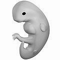
Embryo at 4 weeks after fertilization
-

Fetus at 8 weeks after fertilization
-

Fetus at 18 weeks after fertilization
-
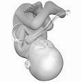
Fetus at 38 weeks after fertilization
-

Relative size in 1st month (simplified illustration)
-

Relative size in 3rd month (simplified illustration)
-

Relative size in 5th month (simplified illustration)
-

Relative size in 9th month (simplified illustration)
One way to observe prenatal development is via ultrasound images. Modern 3D ultrasound images provide greater detail for prenatal diagnosis than the older 2D ultrasound technology. While 3D is popular with parents desiring a prenatal photograph as a keepsake, both 2D and 3D are discouraged by the FDA for non-medical use, but there are no definitive studies linking ultrasound to any adverse medical effects. The following 3D ultrasound images were taken at different stages of pregnancy:
-

75-mm fetus (about 14 weeks gestational age)
-

Fetus at 17 weeks
-

Fetus at 20 weeks
Some people are confused about the differences between an ultrasound and a sonogram. An ultrasound is the actual machine that lets you observe pregnancy. A sonogram is the image of the embryo that the ultrasound produces. 4D Ultrasounds take 3D sonograms. Some people refer to the procedure as prenatal imaging, 3D imaging, a 3D scan, or 4D scan.
Physiological changes

During pregnancy, the woman undergoes many physiological changes, which are entirely normal, including cardiovascular, hematologic, metabolic, renal and respiratory changes that become very important in the event of complications. The body must change its physiological and homeostatic mechanisms in pregnancy to ensure the fetus is provided for. Increases in blood sugar, breathing and cardiac output are all required. Levels of progesterone and oestrogens rise continually throughout pregnancy, suppressing the hypothalamic axis and subsequently the menstrual cycle. The woman and the placenta also produce many hormones.
Management
Prenatal medical care is the medical and nursing care recommended for women before and during pregnancy. The aim of good prenatal care is to detect any potential problems early, to prevent them if possible (through recommendations on adequate nutrition, exercise, vitamin intake etc.), and to direct the woman to appropriate specialists, hospitals, etc. if necessary.
Nutrition
A balanced, nutritious diet is an important aspect of a healthy pregnancy. Eating a healthy diet, balancing carbohydrates, fat, and proteins, and eating a variety of fruits and vegetables, usually ensures good nutrition. Those whose diets are affected by health issues, religious requirements, or ethical beliefs may choose to consult a health professional for specific advice.
Adequate periconceptional folic acid (also called folate or Vitamin B9) intake has been proven to limit fetal neural tube defects, preventing spina bifida, a very serious birth defect. The neural tube develops during the first 28 days of pregnancy, explaining the necessity to guarantee adequate periconceptional folate intake. Folates (from folia, leaf) are abundant in spinach (fresh, frozen, or canned), and are found in green leafy vegetables e.g. salads, beets, broccoli, asparagus, citrus fruits and melons, chickpeas (i.e. in the form of hummus or falafel), and eggs. In the United States and Canada, most wheat products (flour, noodles) are fortified with folic acid.
DHA omega-3 is a major structural fatty acid in the brain and retina, and is naturally found in breast milk. It is important for the woman to consume adequate amounts of DHA during pregnancy and while nursing to support her well-being and the health of her infant. Developing infants cannot produce DHA efficiently, and must receive this vital nutrient from the woman through the placenta during pregnancy and in breast milk after birth.
Several micronutrients are important for the health of the developing fetus, especially in areas of the world where insufficient nutrition is prevalent. In developed areas, such as Western Europe and the United States, certain nutrients such as Vitamin D and calcium, required for bone development, may require supplementation. A 2011 study examined cord blood of healthy neonates and found that low levels of vitamin D are associated with increased risk of lower respiratory tract infection the first year of life.
Dangerous bacteria or parasites may contaminate foods, particularly Listeria and toxoplasma, toxoplasmosis agent. Careful washing of fruits and raw vegetables may remove these pathogens, as may thoroughly cooking leftovers, meat, or processed meat. Soft cheeses may contain Listeria; if milk is raw, the risk may increase. Cat feces pose a particular risk of toxoplasmosis. Pregnant women are also more prone to Salmonella infections from eggs and poultry, which should be thoroughly cooked. Practicing good hygiene in the kitchen can reduce these risks.
Weight gain
Caloric intake must be increased to ensure proper development of the fetus. The amount of weight gained during a single pregnancy varies among women. The Institute of Medicine recommends an overall pregnancy weight gain for women starting pregnancy at a normal weight, with a body mass index of 18.5-24.9, of 25-35 pounds (11.4-15.9 kg). Women who are underweight, with a BMI of less than 18.5, may need to gain between 28-40 lbs. Overweight women are advised to gain between 15-25 lbs, whereas an obese woman may expect to gain between 11-20 lbs. Doctors and dietitians may make different, or more individualized, recommendations for specific patients, based on factors including low maternal age, nutritional status, fetal development, and morbid obesity.
During pregnancy, insufficient or excessive weight gain can compromise the health of the mother and fetus. All women are encouraged to choose a healthy diet regardless of pre-pregnancy weight. Exercise during pregnancy, such as walking and swimming, is recommended for healthy pregnancies. Exercise has notable health benefits for both mother and baby, including preventing excessive weight gain.
Immune tolerance
The fetus inside a pregnant woman may be viewed as an unusually successful allograft, since it genetically differs from the woman. In the same way, many cases of spontaneous abortion may be described in the same way as maternal transplant rejection.
Medication use
Drugs used during pregnancy can have temporary or permanent effects on the fetus. Therefore many physicians would prefer not to prescribe for pregnant women, the major concern being over teratogenicity of the drugs.
Drugs have been classified into categories A,B,C,D and X based on the Food and Drug Administration (FDA) rating system to provide therapeutic guidance based on potential benefits and fetal risks. Drugs like multivitamins that have demonstrated no fetal risks after controlled studies in humans are classified as Category A. On the other hand drugs like thalidomide with proven fetal risks that outweigh all benefits are classified as Category X.
Exposure to toxins
Various toxins pose a significant hazard to fetuses during development. A 2011 study found that virtually all U.S. pregnant women carry multiple chemicals, including some banned since the 1970s, in their bodies. Researchers detected polychlorinated biphenyls, organochlorine pesticides, perfluorinated compounds, phenols, polybrominated diphenyl ethers, phthalates, polycyclic aromatic hydrocarbons, perchlorate PBDEs, compounds used as flame retardants, and dichlorodiphenyltrichloroethane (DDT), a pesticide banned in the United States in 1972, in the bodies of 99 to 100 percent of the pregnant women they tested. Bisphenol A (BPA) was identified in 96 percent of the women surveyed. Several of the chemicals were at the same concentrations that have been associated with negative effects in children from other studies and it is thought that exposure to multiple chemicals can have a greater impact than exposure to only one substance.- Alcohol ingestion during pregnancy may cause fetal alcohol syndrome, a permanent and often devastating birth-defect syndrome. A number of studies have shown that light to moderate drinking during pregnancy might not pose a risk to the fetus, although no amount of alcohol during pregnancy can be guaranteed to be absolutely safe.
- Numerous studies show that children exposed to prenatal cigarette smoke may experience a wide range of behavioral, neurological, and physical difficulties.
- Elemental mercury and methylmercury are two forms of mercury that may pose risks in pregnancy. Methylmercury, a worldwide contaminant of seafood and freshwater fish, is known to produce adverse nervous system effects, especially during brain development. Eating fish is the main source of mercury exposure in humans and some fish may contain enough mercury to harm the developing nervous system of an embryo or fetus, sometimes leading to learning disabilities. Mercury is present in many types of fish, but it is mostly found in certain large fish. The United States Food and Drug Administration and the Environmental Protection Agency advise pregnant women not to eat swordfish, shark, king mackerel and tilefish and limit consumption of albacore tuna to 6 ounces or less a week.
- The Center for Children's Environmental Health reports studies that demonstrate that exposure to air pollution during pregnancy is related to adverse birth outcomes including low birth weight, premature delivery, and heart malformations. Cord blood of exposed babies shows DNA damage that has been linked to cancer. Follow-up studies show a higher level of developmental delays at age three, lower scores on IQ tests and increased behavioral problems at ages six and eight.
- According to the U.S. Centers for Disease Control, the developing nervous system of the fetus is particularly vulnerable to lead toxicity. Neurological toxicity is observed in children of exposed women as a result of the ability of lead to cross the placental barrier and to cause neurological impairment in the fetus. A special concern for pregnant women is that some of the bone lead accumulation is released into the blood during pregnancy. Several studies have provided evidence that even low maternal exposures to lead produce intellectual and behavioral deficits in children1.
- A 2006 study found that children who were exposed prenatally to the insecticide chlorpyrifos had significantly poorer mental and motor development by three years of age and increased risk for behavior problems. A 2007 study using a mouse model suggested that exposure to polycyclic aromatic hydrocarbons prior to conceiving and when lactating reduces the number of eggs in the ovaries of female offspring by two-thirds. A 2009 study of pregnant women exposed to tetrachloroethylene in drinking water found an increased risk of oral clefts and neural tube defects in their children. A 2009 study found that prenatal exposure to phthalates, the chemical compounds used as plasticizers in a wide variety of personal care products, children's toys, and medical devices, may be an environmental risk factor for low birth weight in infants." A 2010 study found that prenatal exposure to flame retardant compounds called polybrominated diphenyl ethers is associated with adverse neurodevelopmental effects in young children.
Sexual activity during pregnancy
Most pregnant women can enjoy sexual activity during pregnancy throughout gravidity. Most research suggests that, during pregnancy, both sexual desire and frequency of sexual relations decrease. In context of this overall decrease in desire, some studies indicate a second-trimester increase, preceding a decrease. However, these decreases are not universal: a significant number of women report greater sexual satisfaction throughout their pregnancies.